
It helps in detecting any tear herniation or. MRI confirms the location of a herniated disc and affected nerves.

Pin On Disc Prolapse Slipped Bulged Disc
MRI scan can diagnose whether the intervertebral disc is herniated.
. How MRI Scans Work. B adults with low back andor leg pain with. This causes the discs to be less able to withstand daily pressures and more susceptible to injuries.
Of course that doesnt mean other potential. Seth Neubardt cervical spine surgical expert tells how the x-ray is used to check the bones in the neck but it cannot reveal a herniated disc. The term slipped disc most commonly refers to a condition known as a herniated.
Your doctor may recommend an x-ray or MRI if your lower back pain resulted from traumatic injury such as a fall or car accident. Myelogram injected dye in your spinal fluid uncovers herniated discs. These images are called three-foot because.
These come in two varieties. During a CT scan a patient lies on a table inside of a tunnel and a. X-rays rule out back pain issues such.
X ray only show pelvic tilt and L5S1 disc space narrowing. To see the disc you must obtain a CT scan or MRI. Unlike an X-ray which can only capture pictures of.
Your doctor may want to request an MRI scan to get clearer pictures of where the herniated disc is located and its severity. Chest X-Ray - Lung disease. Age also plays a role.
MRI scan confirms the location and severity of the slipped disc and helps in finding details of the spinal infection or presence of a possible tumor. A both prospective and retrospective cohort and case-control studies. A herniated disk also can give you a feeling of tingling or numbness.
MRI actually show L34 disc herniation red arrow no L5S1 disc herniation yellow arrow that was suspected in X ray. Patients who complain of experiencing symptoms related to herniated discs. We applied the following selection criteria.
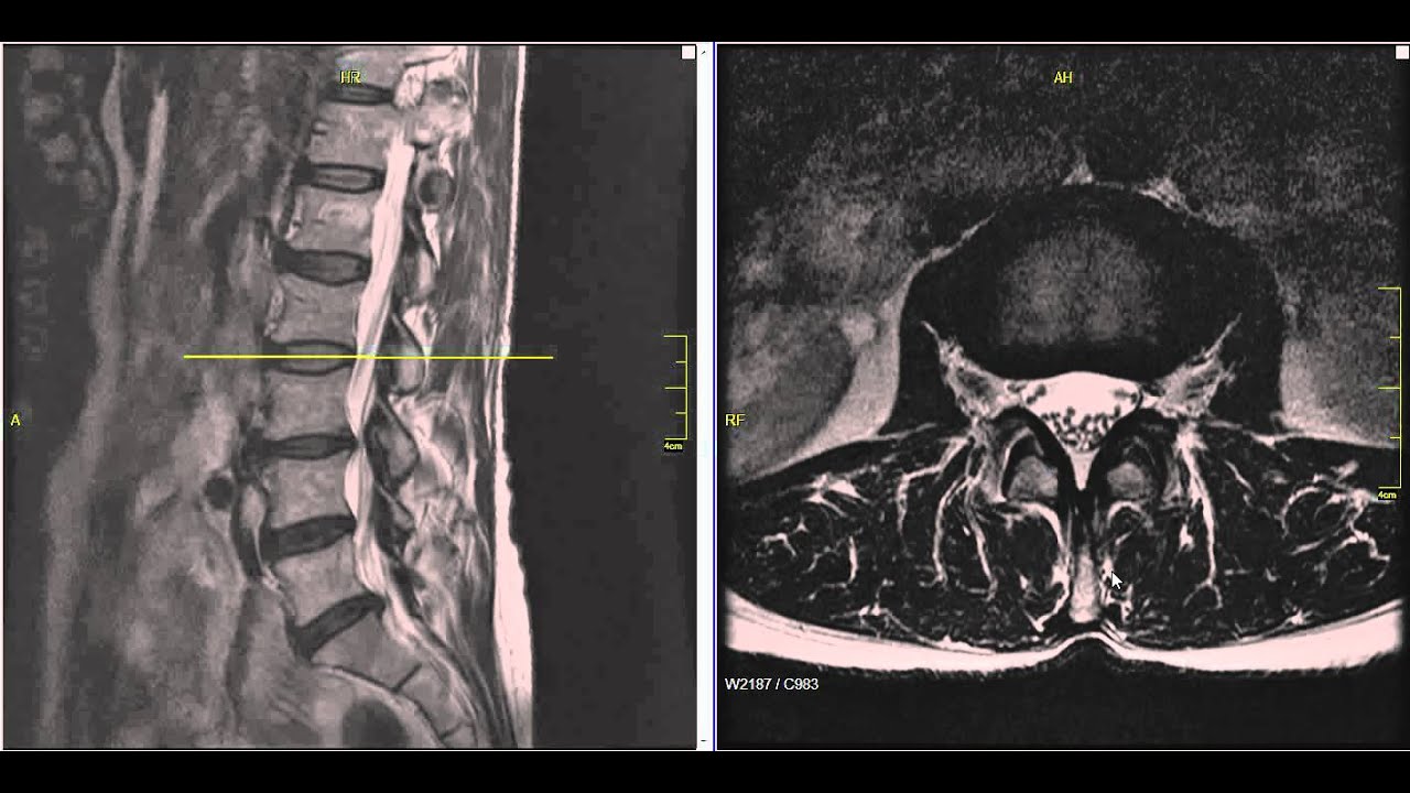
In an MRI image the intervertebral disc is shown as white due to the fact that it contains water. Plain X-rays dont detect herniated disks but they can rule out other causes of back pain such as an infection tumor spinal alignment issues or a broken bone. An MRI scan takes longer than.
Diagnostic imaging including MRI CT and X-Ray is used to diagnose a range of spinal conditions from mild to severe. Anteriorposterior X-rays which are taken from the front and then the back and lateral X-rays taken from the side. Chest X-Ray - Basic Interpretation.
If you would like us to review your MRI or X-rays at no cost to see if we might be able to help you with true spinal decompression using our 13 year proven protocol with the. The MRI will then take a picture. The doctor can use an X-ray to look for pressure on your nerves and spine by injecting a dye into your body.
Obtaining an MRI magnetic resonance imaging scan can be a very important step in correctly diagnosing a herniated disc in your spine. Undergo an MRI to. Chest X-ray - Heart Failure.
These strong components will make the protons in the affected area line up. Herniated discs will not show up on an X-ray. An x-ray CT scan or MRI scan may be ordered to test for herniated disc in neck or any other region on the spine where the patient complains of pain.
MRI scans of herniated discs are a helpful test used to determine the exact location and condition of the problem. Spinal abnormalities such as herniated discs are. As you get older your disks tend to break down.
Disc herniation refers to the displacement of intervertebral disc material beyond the normal confines of the disc but involving less than 25 of the circumference to distinguish. Unlike an x-ray an MRI uses radio waves and magnetic fields to produce images. A normal disc will show as white whereas a degenerated disc or herniated.
The MRI protocol for examination of the. Amongst the available imaging. It provides higher quality images than an X-ray and can be used to diagnose a variety of health conditions.
The affected part of your back may also feel weak.

Pin On Resonancia Ortopedia

Pin On Anatomy

Lateral Ct Scan Shows A Herniated Disc Protruding Into The Spinal Canal Cauda Equina Syndrome Cauda Equina Diagnostic Imaging

Cervical X Ray Vs Mri Disc Herniation Disk Herniation Mri Subluxation

Stem Cells For Degenerative Disc Disease Ddd Therapy Http Stemcellthailand Org Therapies Degener Degenerative Disc Disease Degenerative Disc Medical Anatomy

Pin On Anatomy

Normal Mri Lumbar Medical Anatomy Radiology Imaging Medical Knowledge

Neck And Back Degenerative Spondylolisthesis Conditions Spondylolisthesis Spinal Canal Spine Surgery
Lumbar Disc Herniation Mri Explained Dr Jeffrey P Johnson Hd Youtube Lumbar Disc Disk Herniation Mri

Pin On Back

Pin On Mri

Pin On Conditions
![]()
Lumbar Spine Mri Unidad Especializada En Ortopedia Y Traumatologia En Bogota Colombia Pbx 6923370 Www Unidadortopedia Com Nuclear Medicine Mri Radiology

Disc Herniation Best Ways To Help Your Lumbar Disc Herniation Degenerative Disc Degenerative Disc Disease Bulging Disc

Pin On Bulging Disc

Cauda Equina Syndrome From Lumbar Disc Herniation Cauda Equina Cauda Equina Syndrome Lumbar Disc

Prolapsed Cervical Disk Intervertebral Disc Pathology Nucleus Pulpusus Bulges Out Into A Weakend Area Of Annulus Fibr Herniated Disc Herniated Cervical Disc

Pin On Hernie

T2 Mri Lumbar Spine Sagittal View L4 5 Sever Disc Herniation With Spinal Stenosis Disc Degeneration At L4 L5 Also Lumbar Lordosis Spinal Stenosis Stenosis


